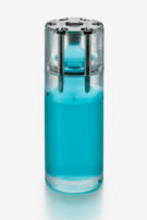
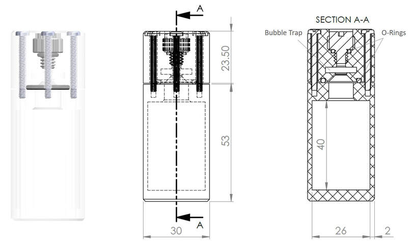
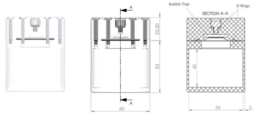
Phantech’s uniformity phantoms are designed to perform quality control and calibration on field uniformity, normalization and quantification calibration scans in nuclear medicine. They are constructed to ensure a perfectly homogenous concentration by minimizing air bubbles and preventing sticking to the interior surface. Configured in two sizes to simulate typical rodent sizes (ie. mice and rats), these uniformity phantoms will allow one to customize protocols and calibrations in order to yield the highest quantitative accuracy for each scanning condition. Each configuration incorporates several design features that will make the job easier, including two o-ring sealed openings (large one for filling milliliters and a smaller one for filling microliters), a bubble trap to ensure that any bubbles will not be in the uniformity region, and nested fasteners to maintain a uniform diameter across the phantom.
See below for more info and contact us today for pricing info!
See below for more info and contact us today for pricing info!
Available Sizes
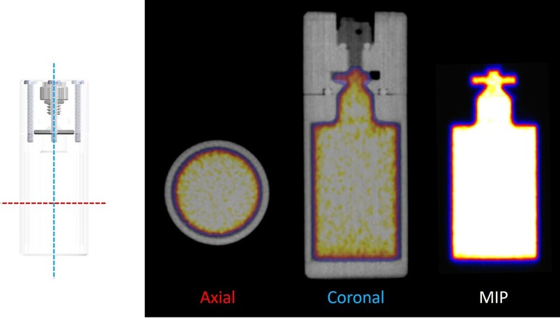
30 mm OD (26 mm ID) - Small
CAD drawings and dimensions (units in mm)
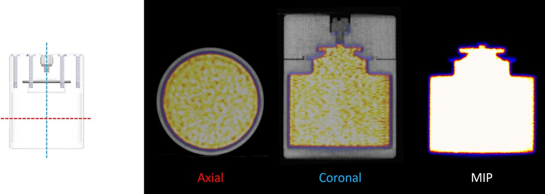
PET Images (F-18)
60 mm OD (56 mm ID) - Large
CAD drawings and dimensions (units in mm)
PET Images (F-18)
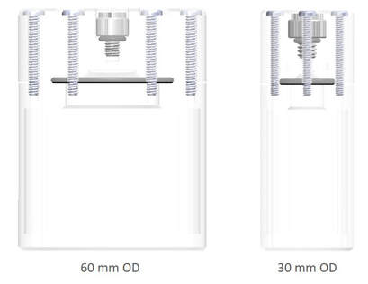
Size Comparison
Additional Info
What are normalization, uniformity, and quantification calibration scans and why do I need them?
Normalization
Your PET scanner detector ring is made up of many independent modular parts. At the top level, your PET ring consists of dozens, or more, of detector blocks that are made up of an array of imperfect scintillation crystals that are coupled to a photomultiplier tube. In all, your PET rings are made of tens to hundreds of detectors with slight differences in geometry and electronic noise, thousands of crystals with varying efficiencies, and hundreds of photomultiplier tubes with variable gains. If crystal production was perfect and detector electronics were stable, when scanning a phantom with a known uniform activity concentration, each crystal pair on a line of response should yield the exact same signal. Since this is not the case, you must apply a correction factor to each PET reconstruction that is based on the discrepancy between what the detector pairs measured in the uniformity phantom and what was actually scanned in the uniformity phantom.
In other words, a “normalization” corrects for detector pair inefficiencies resulting in a balanced response of all detector pairs. It is important to note that a new normalization is required whenever there is a change in scanner parameter, e.g. energy window, timing window, span, ring difference, etc. Not applying a normalization scan can lead to artifacts and poor counting statistics. The general procedure for normalization scans involves filling Phantech’s rat uniformity phantom with a known activity (typically F-18) concentration and scan in the center of the PET field-of-view (FOV). The rat uniformity phantom is ideal for us in preclinical systems because it covers most of the FOV which is important for generating lines of response throughout the field.
In other words, a “normalization” corrects for detector pair inefficiencies resulting in a balanced response of all detector pairs. It is important to note that a new normalization is required whenever there is a change in scanner parameter, e.g. energy window, timing window, span, ring difference, etc. Not applying a normalization scan can lead to artifacts and poor counting statistics. The general procedure for normalization scans involves filling Phantech’s rat uniformity phantom with a known activity (typically F-18) concentration and scan in the center of the PET field-of-view (FOV). The rat uniformity phantom is ideal for us in preclinical systems because it covers most of the FOV which is important for generating lines of response throughout the field.
Quantification Calibration
After your system is normalized, it is necessary to perform a quantification calibration of your scanner for every combination of radioisotope, reconstruction type, parameters (e.g. numbers of iterations and subsets, filter types), and application of attenuation and scatter correction. Every scanner’s energy window is configured so that the peak energy of the 511 keV gammas produced from the annihilation of a positron is detected. An energy window must exist in order to detect the distribution of photon energies as a result of Compton scatter while also achieving significant count rates. Unfortunately, radioisotopes have other characteristic gamma emissions that fall within this window and add noise. Therefore, every combination of PET radioisotope and reconstruction will need to be corrected with a respective calibration factor. If done properly, the quantification calibration will allow you to assign concentration units, such as microcuries per milliliter, to each voxel value so you can accurately calculate an activity concentration based on an injected amount of activity. Without a quantification calibration, you cannot measure an accurate activity concentration for a given radioisotope and reconstruction of your PET image.
The general procedure for quantification calibration scans involves filling Phantech’s uniformity phantom that most closely resembles the size of the scan subject with a known activity concentration. The phantom is then scanned in the center of the PET field-of-view (FOV) with 5-10x more counts than a typical PET acquisition protocol. A calibration factor is then calculated based on the difference between the known activity concentration in the phantom versus what was detected in the PET scan.
The general procedure for quantification calibration scans involves filling Phantech’s uniformity phantom that most closely resembles the size of the scan subject with a known activity concentration. The phantom is then scanned in the center of the PET field-of-view (FOV) with 5-10x more counts than a typical PET acquisition protocol. A calibration factor is then calculated based on the difference between the known activity concentration in the phantom versus what was detected in the PET scan.
Image Field Uniformity
Image field uniformity is a quality control procedure that should be performed routinely. If your system is correctly normalized and quantification calibrated, then the image field should be uniform. That is, the measured radioactivity throughout the entire PET FOV should accurately measure the activity concentration in the uniformity phantom.
The procedure for measuring image field uniformity requires scanning the uniformity phantom (typically F-18) in the center of the FOV. The mean and the standard deviation of the voxels inside the phantom are then calculated for QC assessment.
The procedure for measuring image field uniformity requires scanning the uniformity phantom (typically F-18) in the center of the FOV. The mean and the standard deviation of the voxels inside the phantom are then calculated for QC assessment.
