
|

|
Key features
• Phantech’s linear-filling technology
-~30 second to fill
-Luer lock fittings ensure no spilling
-No bubbles
-Videos provided for step-by-step guidance
• Spheres simulate rodent tumors, organs and tissues
• Automatic segmentation and analysis with Imalytics (Gremse-IT) Preclinical Software
-Recovery coefficient curves (RCC) generated with the click of a button
-Apply PVC to in vivo ROIs using RCCs
• 3 standard versions with varying sphere sizes (27, 34, 49mm OD)
• Fillable warm background to simulate spill-in/background
• Internal reference/normalization volume to accommodate sites that do not have tools to measure absolute activity concentration
• Sealed sources (68Ge, 22Na) available
• Custom sizing available
• Compatible with most imaging systems and modalities, including MRI
Description & Applications
Phantech’s Partial Volume Correction (PVC) phantoms are used to generate recovery coefficient (RC) calibration curves. By comparing the measured activity concentration in each sphere to the known activity concentration, one can characterize the signal recovered as a function of sphere size (simulating tumors & organs) by an imaging system and then apply a correction factor. Quantitative accuracy is critical for molecular imaging research and theranostic personalized medicine.
Designed with one continuous channel connecting numerous precise spherical or ellipsoidal volumes, Phantech's PVC phantoms allow for quick, easy and safe filling.
Common applications:
Additional Information
What are Partial Volume Effects (PVEs)?
PVEs are an inherent physical phenomenon present in all imaging systems largely due to sampling, blurring, motion, and physical hardware constraints. However, in nuclear medicine (particularly with PET and SPECT imaging), PVEs are compounded by a radioisotope’s finite positron emission energy and its respective inherent mean positron range. For a radioisotope with a relatively short positron range (ie. 18F in a static system, the intrinsic spatial resolution of the PET scanner will have a greater influence on partial volume effects (PVEs) than the positron range of the radioisotope. On the other hand, radioisotopes with relatively long positron ranges (ie. 124I, 89Zr) are largely responsible for PVEs due to the displacement of the originating positron emission voxel to the detected annihilation voxel. In either case, PVEs play a major role in the precision and accuracy of quantitative PET data, particularly with the degradation of measured radiotracer concentration in smaller structures using radioisotopes with long positron ranges.
Why do I need to correct for PVEs?
PVEs contribute to imprecise and inaccurate quantitative data in ALL of the approximately 2 million clinical positron emission tomography (PET) and ~14.5 million single-photon emission computed tomography (SPECT) scans per year, and tens of millions of preclinical PET and SPECT scans per year. Clinically, not correcting for PVEs can have serious negative consequences on patient management and outcomes, including but not limited to, progressive disease, radiotoxicity of normal tissue in theranostic applications, erroneous staging and prognosis, and the inability to compare data in multi-site trials, which collectively contribute to increased healthcare costs. Preclinically, not correcting for PVEs can have serious negative consequences as well, including but not limited to, erroneous data interpretation and conclusions, radiotoxicity of normal tissue in theranostic applications, and the inability to compare data between institutions.
Designed with one continuous channel connecting numerous precise spherical or ellipsoidal volumes, Phantech's PVC phantoms allow for quick, easy and safe filling.
Common applications:
- Recovery coefficient measurements
- Partial volume correction for dosimetry
- Co-registration (PET, SPECT, CT, optical, MRI, X-ray, MPI)
- Reconstruction optimization
- Multi-modality applications
- Standardization
- Image quality assessment
- Training
- Marketing
Additional Information
What are Partial Volume Effects (PVEs)?
PVEs are an inherent physical phenomenon present in all imaging systems largely due to sampling, blurring, motion, and physical hardware constraints. However, in nuclear medicine (particularly with PET and SPECT imaging), PVEs are compounded by a radioisotope’s finite positron emission energy and its respective inherent mean positron range. For a radioisotope with a relatively short positron range (ie. 18F in a static system, the intrinsic spatial resolution of the PET scanner will have a greater influence on partial volume effects (PVEs) than the positron range of the radioisotope. On the other hand, radioisotopes with relatively long positron ranges (ie. 124I, 89Zr) are largely responsible for PVEs due to the displacement of the originating positron emission voxel to the detected annihilation voxel. In either case, PVEs play a major role in the precision and accuracy of quantitative PET data, particularly with the degradation of measured radiotracer concentration in smaller structures using radioisotopes with long positron ranges.
Why do I need to correct for PVEs?
PVEs contribute to imprecise and inaccurate quantitative data in ALL of the approximately 2 million clinical positron emission tomography (PET) and ~14.5 million single-photon emission computed tomography (SPECT) scans per year, and tens of millions of preclinical PET and SPECT scans per year. Clinically, not correcting for PVEs can have serious negative consequences on patient management and outcomes, including but not limited to, progressive disease, radiotoxicity of normal tissue in theranostic applications, erroneous staging and prognosis, and the inability to compare data in multi-site trials, which collectively contribute to increased healthcare costs. Preclinically, not correcting for PVEs can have serious negative consequences as well, including but not limited to, erroneous data interpretation and conclusions, radiotoxicity of normal tissue in theranostic applications, and the inability to compare data between institutions.
Available sizes
Our PVC phantoms are available in 3 standard sizes: 27, 34 and 49mm outside diameter. Customization is possible!
|

|
*Volumes match the figure above, including sphere void chain (purple), reference volume (green), and background (blue)
Customization
Yes, we can customize our PVC phantoms. Customization options include a full 360 ̊ or half 180 ̊ sleeve for a "warm" spill-in compartment, a second channel of "micro-spheres", and additional ellipsoidal PVC phantoms with varying aspect ratios (i.e. 1:2 & 2:3) to create a comprehensive 3D recovery coefficient correction curve.
Contact us at [email protected] for custom requests!
Contact us at [email protected] for custom requests!
image and video gallery
See this video showing the filling of Phantech's Partial Volume Correction (PVC) phantom in <1 minute with no air bubbles!

CAD rendering (left) of the 34 mm OD PVC Phantom, next to a CAD rendering (right) of the three fillable volumes
|

Computer-aided design (CAD) rendering of our 34 mm OD PVC phantom, including a front view (left) and a side view (right)
|

F18 PET image (left), Ga68 PET image (center), and a CAD rendering of the spherical voids in the PVC27
|
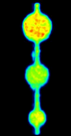
microSPECT scan of a single concentration of 177Lu using Phantech's 27mm OD PVC phantom
|

Recovery coefficients as a function of sphere diameter for the same radioisotope using various reconstruction algorithms.
SPecificiation sheet
| Phantech_PVC_specsheet.pdf |
INNOVATIVE DESIGN
|
Phantech's PVC phantoms come standard with a large internal reference calibration void that is "100%" recovered, and each sphere can be normalized to this reference void. This internal calibration method allows for a more robust and standardized method of generating RC curves because the RC accuracy is not dependent on the accuracy of another calibration device. Alternatively, RCs can be generated by comparing the activity concentration of the spheres to the true activity concentration. Phantech's PVC phantoms also contain more measurements points (spheres) than any other PVC phantoms on the market to create the most accurate RC curves. |

Example micro-PVC phantom with an outer diameter (OD) of 27 mm. Three separately fillable volumes, include a chain of spherical voids, a reference volume, and a warm background.
|
AUTOMATED ANALYSIS WITH IMALYTICS PRECLINICAL (GREMSE-IT)
Watch this short video showing the quick filling of Phantech's Partial Volume Correction (PVC) Phantom and automated analysis using Imalytics (Gremse-IT)
|
Automated Analysis Workflow
1. Fill micro PVC phantom 2. Perform imaging scan 3. Load image file into Imalytics 4. Select the phantom from the dropdown menu 5. Generate and save report |

|
ORDER!
Lead time: <2 weeks.
Email [email protected] for quotes, custom requests, and sealed sources (68Ge, 22Na)
Email [email protected] for quotes, custom requests, and sealed sources (68Ge, 22Na)

Madison, WI
www.phantechmedical.com
www.phantechmedical.com